- Главная
- Разное
- Образование
- Спорт
- Естествознание
- Природоведение
- Религиоведение
- Французский язык
- Черчение
- Английский язык
- Астрономия
- Алгебра
- Биология
- География
- Геометрия
- Детские презентации
- Информатика
- История
- Литература
- Математика
- Музыка
- МХК
- Немецкий язык
- ОБЖ
- Обществознание
- Окружающий мир
- Педагогика
- Русский язык
- Технология
- Физика
- Философия
- Химия
- Шаблоны, фоны, картинки для презентаций
- Экология
- Экономика
Презентация, доклад по дисциплине Английский язык на тему Kidney (ГБОУ НО НМК)
Содержание
- 1. Презентация по дисциплине Английский язык на тему Kidney (ГБОУ НО НМК)
- 2. KidneyThe kidneys are two bean-shaped organs found
- 3. Слайд 3
- 4. NephronThe nephron is the structural and functional
- 5. Renal physiology is the study of kidney
- 6. Procedures used in the management of kidney
- 7. Renal pyramid Interlobular artery Renal artery Renal
- 8. HydronephrosisHydronephrosis—literally "water inside the kidney"—refers to distension
- 9. Signs and symptomsThe signs and symptoms of
- 10. PyelonephritisPyelonephritis is inflammation of the kidney, typically
- 11. Signs and symptomsSigns and symptoms of acute
- 12. GlomerulonephritisGlomerulonephritis (GN), also known as glomerular nephritis,
- 13. As it is not strictly a single
- 14. Renal cell carcinomaRenal cell carcinoma (RCC) is
- 15. Signs and symptomsHistorically, medical practitioners expected a
- 16. Thank you
KidneyThe kidneys are two bean-shaped organs found on the left and right sides of the body in vertebrates. They are located at the back of the abdominal cavity in the retroperitoneal space. In adults they are
Слайд 2Kidney
The kidneys are two bean-shaped organs found on the left and
right sides of the body in vertebrates. They are located at the back of the abdominal cavity in the retroperitoneal space. In adults they are about 11 centimetres (4.3 in) in length. They receive blood from the paired renal arteries; blood exits into the paired renal veins. Each kidney is attached to a ureter, a tube that carries excreted urine to the bladder.
Слайд 4Nephron
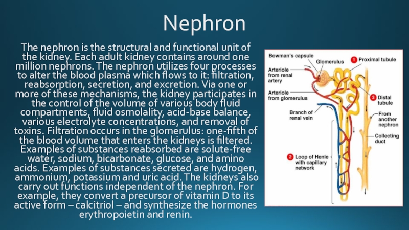
The nephron is the structural and functional unit of the kidney.
Each adult kidney contains around one million nephrons. The nephron utilizes four processes to alter the blood plasma which flows to it: filtration, reabsorption, secretion, and excretion. Via one or more of these mechanisms, the kidney participates in the control of the volume of various body fluid compartments, fluid osmolality, acid-base balance, various electrolyte concentrations, and removal of toxins. Filtration occurs in the glomerulus: one-fifth of the blood volume that enters the kidneys is filtered. Examples of substances reabsorbed are solute-free water, sodium, bicarbonate, glucose, and amino acids. Examples of substances secreted are hydrogen, ammonium, potassium and uric acid. The kidneys also carry out functions independent of the nephron. For example, they convert a precursor of vitamin D to its active form – calcitriol – and synthesize the hormones erythropoietin and renin.
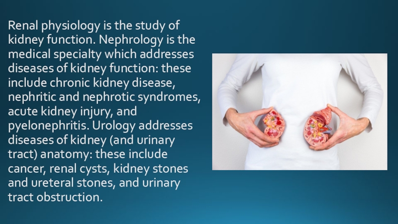
Слайд 5Renal physiology is the study of kidney function. Nephrology is the
medical specialty which addresses diseases of kidney function: these include chronic kidney disease, nephritic and nephrotic syndromes, acute kidney injury, and pyelonephritis. Urology addresses diseases of kidney (and urinary tract) anatomy: these include cancer, renal cysts, kidney stones and ureteral stones, and urinary tract obstruction.
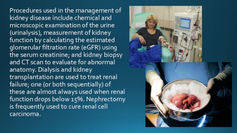
Слайд 6Procedures used in the management of kidney disease include chemical and
microscopic examination of the urine (urinalysis), measurement of kidney function by calculating the estimated glomerular filtration rate (eGFR) using the serum creatinine; and kidney biopsy and CT scan to evaluate for abnormal anatomy. Dialysis and kidney transplantation are used to treat renal failure; one (or both sequentially) of these are almost always used when renal function drops below 15%. Nephrectomy is frequently used to cure renal cell carcinoma.
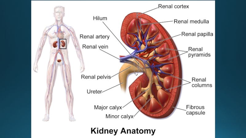
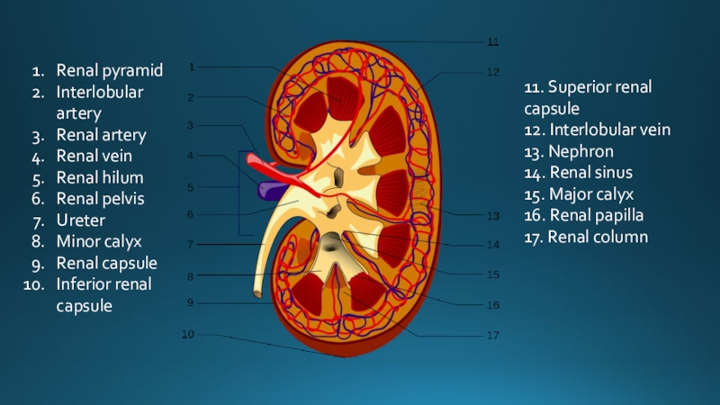
Слайд 7Renal pyramid
Interlobular artery
Renal artery
Renal vein
Renal hilum
Renal
pelvis
Ureter
Minor calyx
Renal capsule
Inferior renal capsule
Ureter
Minor calyx
Renal capsule
Inferior renal capsule
11. Superior renal capsule
12. Interlobular vein
13. Nephron
14. Renal sinus
15. Major calyx
16. Renal papilla
17. Renal column
Слайд 8Hydronephrosis
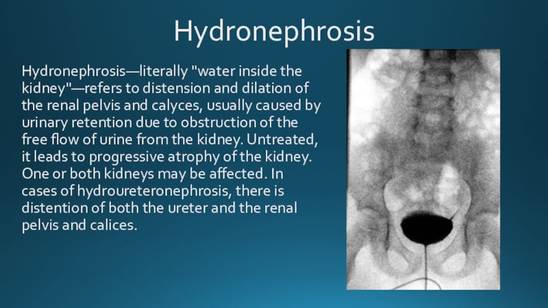
Hydronephrosis—literally "water inside the kidney"—refers to distension and dilation of the
renal pelvis and calyces, usually caused by urinary retention due to obstruction of the free flow of urine from the kidney. Untreated, it leads to progressive atrophy of the kidney. One or both kidneys may be affected. In cases of hydroureteronephrosis, there is distention of both the ureter and the renal pelvis and calices.
Слайд 9Signs and symptoms
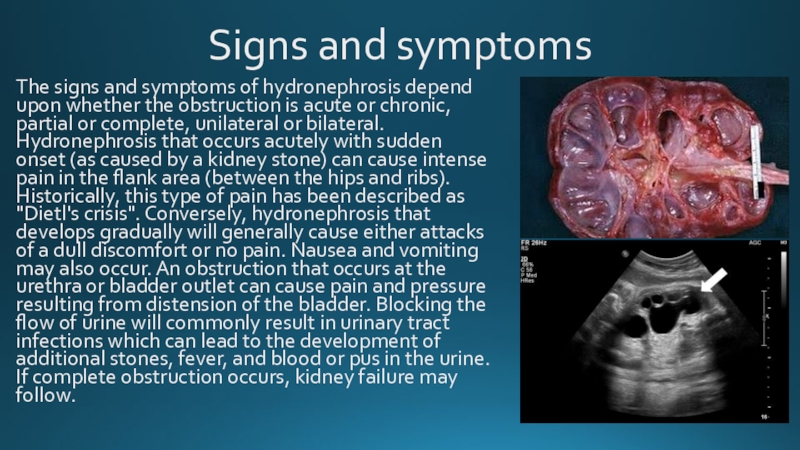
The signs and symptoms of hydronephrosis depend upon whether
the obstruction is acute or chronic, partial or complete, unilateral or bilateral. Hydronephrosis that occurs acutely with sudden onset (as caused by a kidney stone) can cause intense pain in the flank area (between the hips and ribs). Historically, this type of pain has been described as "Dietl's crisis". Conversely, hydronephrosis that develops gradually will generally cause either attacks of a dull discomfort or no pain. Nausea and vomiting may also occur. An obstruction that occurs at the urethra or bladder outlet can cause pain and pressure resulting from distension of the bladder. Blocking the flow of urine will commonly result in urinary tract infections which can lead to the development of additional stones, fever, and blood or pus in the urine. If complete obstruction occurs, kidney failure may follow.
Слайд 10Pyelonephritis
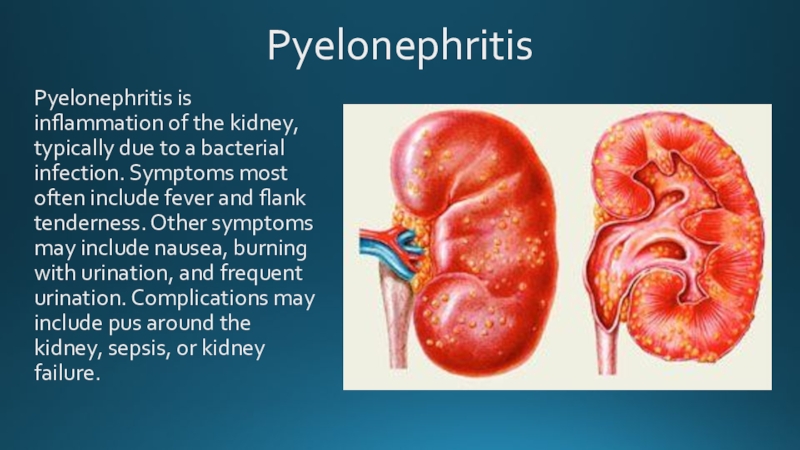
Pyelonephritis is inflammation of the kidney, typically due to a bacterial
infection. Symptoms most often include fever and flank tenderness. Other symptoms may include nausea, burning with urination, and frequent urination. Complications may include pus around the kidney, sepsis, or kidney failure.
Слайд 11Signs and symptoms
Signs and symptoms of acute pyelonephritis generally develop rapidly
over a few hours or a day. It can cause high fever, pain on passing urine, and abdominal pain that radiates along the flank towards the back. There is often associated vomiting.
Chronic pyelonephritis causes persistent flank or abdominal pain, signs of infection (fever, unintentional weight loss, malaise, decreased appetite), lower urinary tract symptoms and blood in the urine. Chronic pyelonephritis can in addition cause fever of unknown origin. Furthermore, inflammation-related proteins can accumulate in organs and cause the condition AA amyloidosis.
Chronic pyelonephritis causes persistent flank or abdominal pain, signs of infection (fever, unintentional weight loss, malaise, decreased appetite), lower urinary tract symptoms and blood in the urine. Chronic pyelonephritis can in addition cause fever of unknown origin. Furthermore, inflammation-related proteins can accumulate in organs and cause the condition AA amyloidosis.
Слайд 12Glomerulonephritis
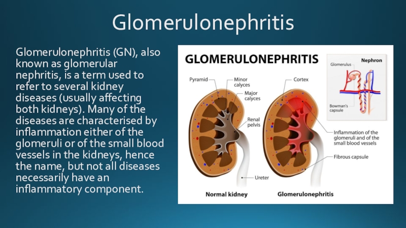
Glomerulonephritis (GN), also known as glomerular nephritis, is a term used
to refer to several kidney diseases (usually affecting both kidneys). Many of the diseases are characterised by inflammation either of the glomeruli or of the small blood vessels in the kidneys, hence the name, but not all diseases necessarily have an inflammatory component.
Слайд 13As it is not strictly a single disease, its presentation depends
on the specific disease entity: it may present with isolated hematuria and/or proteinuria (blood or protein in the urine); or as a nephrotic syndrome, a nephritic syndrome, acute kidney injury, or chronic kidney disease.
They are categorized into several different pathological patterns, which are broadly grouped into non-proliferative or proliferative types. Diagnosing the pattern of GN is important because the outcome and treatment differs in different types. Primary causes are intrinsic to the kidney. Secondary causes are associated with certain infections (bacterial, viral or parasitic pathogens), drugs, systemic disorders (SLE, vasculitis), or diabetes.
They are categorized into several different pathological patterns, which are broadly grouped into non-proliferative or proliferative types. Diagnosing the pattern of GN is important because the outcome and treatment differs in different types. Primary causes are intrinsic to the kidney. Secondary causes are associated with certain infections (bacterial, viral or parasitic pathogens), drugs, systemic disorders (SLE, vasculitis), or diabetes.
Слайд 14Renal cell carcinoma
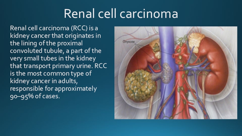
Renal cell carcinoma (RCC) is a kidney cancer that
originates in the lining of the proximal convoluted tubule, a part of the very small tubes in the kidney that transport primary urine. RCC is the most common type of kidney cancer in adults, responsible for approximately 90–95% of cases.
Слайд 15Signs and symptoms
Historically, medical practitioners expected a person to present with
three findings. This classic triad is 1: haematuria, which is when there is blood present in the urine, 2: flank pain, which is pain on the side of the body between the hip and ribs, and 3: an abdominal mass, similar to bloating but larger. It is now known that this classic triad of symptoms only occurs in 10–15% of cases, and is usually indicative that the renal cell carcinoma (RCC) is in an advanced stage.[7] Today, RCC is often asymptomatic (meaning few to no symptoms) and is generally detected incidentally when a person is being examined for other ailments.
Other signs and symptom may include haematuria; loin pain; abdominal mass; malaise, which is a general feeling of unwellness; weight loss and/or loss of appetite; anaemia resulting from depression of erythropoietin; erythrocytosis (increased production of red blood cells) due to increased erythropoietin secretion; varicocele, which is seen in males as an enlargement of the pampiniform plexus of veins draining the testis (more often the left testis) hypertension (high blood pressure) resulting from secretion of renin by the tumour; hypercalcemia, which is elevation of calcium levels in the blood; sleep disturbance or night sweats; recurrent fevers;[9] and chronic fatigue.
Other signs and symptom may include haematuria; loin pain; abdominal mass; malaise, which is a general feeling of unwellness; weight loss and/or loss of appetite; anaemia resulting from depression of erythropoietin; erythrocytosis (increased production of red blood cells) due to increased erythropoietin secretion; varicocele, which is seen in males as an enlargement of the pampiniform plexus of veins draining the testis (more often the left testis) hypertension (high blood pressure) resulting from secretion of renin by the tumour; hypercalcemia, which is elevation of calcium levels in the blood; sleep disturbance or night sweats; recurrent fevers;[9] and chronic fatigue.
