г.
- Главная
- Разное
- Образование
- Спорт
- Естествознание
- Природоведение
- Религиоведение
- Французский язык
- Черчение
- Английский язык
- Астрономия
- Алгебра
- Биология
- География
- Геометрия
- Детские презентации
- Информатика
- История
- Литература
- Математика
- Музыка
- МХК
- Немецкий язык
- ОБЖ
- Обществознание
- Окружающий мир
- Педагогика
- Русский язык
- Технология
- Физика
- Философия
- Химия
- Шаблоны, фоны, картинки для презентаций
- Экология
- Экономика
Презентация, доклад по английскому языку на тему Innovations in burn treatment
Содержание
- 1. Презентация по английскому языку на тему Innovations in burn treatment
- 2. BurnA burn is a type of injury
- 3. CauseBurns are caused by a variety of
- 4. Слайд 4
- 5. Kind of burnsThermal:In the United States, fire
- 6. Слайд 6
- 7. ChemicalChemicals cause from 2 to 11% of
- 8. Слайд 8
- 9. ElectricalElectrical burns or injuries are classified as
- 10. Слайд 10
- 11. RadiationRadiation burns may be caused by protracted
- 12. Слайд 12
- 13. Non accidentalIn those hospitalized from scalds or
- 14. Innovations in burn treatmentTechnology the treatment of
- 15. Слайд 15
- 16. SPRAY-ON SKINAviata Medical’s ReCell device creates what
- 17. While test-tube skin can help when there’s
- 18. (a) An illustration of a microfluidic platform
- 19. The process begins with stem cells that
- 20. PREVENTING BURN SCARS
- 21. Indeed, one reason burns are particularly difficult
- 22. MONITORING HEALINGAn epidermal blood-flow sensor and (b)
- 23. Flexible electronics are the name of the
- 24. Thanks for attention
BurnA burn is a type of injury to skin, or other tissues, caused by heat, cold, electricity, chemicals, friction, or radiation. Most burns are due to heat from hot liquids, solids, or fire. Females in many
Слайд 2Burn
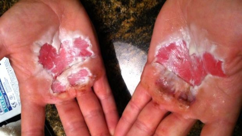
A burn is a type of injury to skin, or other
tissues, caused by heat, cold, electricity, chemicals, friction, or radiation. Most burns are due to heat from hot liquids, solids, or fire. Females in many areas of the world have a higher risk related to the more frequent use of open cooking fires or unsafe cook stoves. Alcoholism and smoking are other risk factors. Burns can also occur as a result of self harm or violence between people.
Слайд 3Cause
Burns are caused by a variety of external sources classified as
thermal (heat-related), chemical, electrical, and radiation. In the United States, the most common causes of burns are: fire or flame (44%), scalds (33%), hot objects (9%), electricity (4%), and chemicals (3%). Most (69%) burn injuries occur at home or at work (9%), and most are accidental, with 2% due to assault by another, and 1-2% resulting from a suicide attempt. These sources can cause inhalation injury to the airway and/or lungs, occurring in about 6%.
Слайд 5Kind of burns
Thermal:
In the United States, fire and hot liquids are
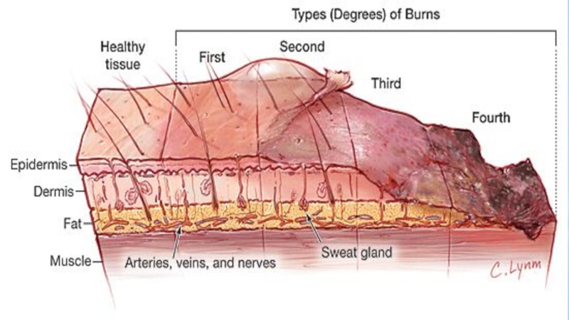
the most common causes of burns. Of house fires that result in death, smoking causes 25% and heating devices cause 22%. Almost half of injuries are due to efforts to fight a fire. Scalding is caused by hot liquids or gases and most commonly occurs from exposure to hot drinks, high temperature tap water in baths or showers, hot cooking oil, or steam. Scald injuries are most common in children under the age of five and, in the United States and Australia, this population makes up about two-thirds of all burns. Contact with hot objects is the cause of about 20-30% of burns in children. Generally, scalds are first- or second-degree burns, but third-degree burns may also result, especially with prolonged contact. Fireworks are a common cause of burns during holiday seasons in many countries. This is a particular risk for adolescent males.
Слайд 7Chemical
Chemicals cause from 2 to 11% of all burns and contribute
to as many as 30% of burn-related deaths. Chemical burns can be caused by over 25,000 substances, most of which are either a strong base (55%) or a strong acid (26%). Most chemical burn deaths are secondary to ingestion. Common agents include: sulfuric acid as found in toilet cleaners, sodium hypochlorite as found in bleach, and halogenated hydrocarbons as found in paint remover, among others. Hydrofluoric acid can cause particularly deep burns that may not become symptomatic until some time after exposure. Formic acid may cause the breakdown of significant numbers of red blood cells.
Слайд 9Electrical
Electrical burns or injuries are classified as high voltage (greater than
or equal to 1000 volts), low voltage (less than 1000 volts), or as flash burns secondary to an electric arc. The most common causes of electrical burns in children are electrical cords (60%) followed by electrical outlets (14%). Lightning may also result in electrical burns. Risk factors for being struck include involvement in outdoor activities such as mountain climbing, golf and field sports, and working outside. Mortality from a lightning strike is about 10%.
While electrical injuries primarily result in burns, they may also cause fractures or dislocations secondary to blunt force trauma or muscle contractions. In high voltage injuries, most damage may occur internally and thus the extent of the injury cannot be judged by examination of the skin alone. Contact with either low voltage or high voltage may produce cardiac arrhythmias or cardiac arrest.
While electrical injuries primarily result in burns, they may also cause fractures or dislocations secondary to blunt force trauma or muscle contractions. In high voltage injuries, most damage may occur internally and thus the extent of the injury cannot be judged by examination of the skin alone. Contact with either low voltage or high voltage may produce cardiac arrhythmias or cardiac arrest.
Слайд 11Radiation
Radiation burns may be caused by protracted exposure to ultraviolet light
(such as from the sun, tanning booths or arc welding) or from ionizing radiation (such as from radiation therapy, X-rays or radioactive fallout). Sun exposure is the most common cause of radiation burns and the most common cause of superficial burns overall. There is significant variation in how easily people sunburn based on their skin type. Skin effects from ionizing radiation depend on the amount of exposure to the area, with hair loss seen after 3 Gy, redness seen after 10 Gy, wet skin peeling after 20 Gy, and necrosis after 30 Gy. Redness, if it occurs, may not appear until some time after exposure. Radiation burns are treated the same as other burns. Microwave burns occur via thermal heating caused by the microwaves. While exposures as short as two seconds may cause injury, overall this is an uncommon occurrence.
Слайд 13Non accidental
In those hospitalized from scalds or fire burns, 3–10% are
from assault. Reasons include: child abuse, personal disputes, spousal abuse, elder abuse, and business disputes. An immersion injury or immersion scald may indicate child abuse. It is created when an extremity or the lower body (buttock or perineum) is held under the surface of hot water. It typically produces a sharp upper border and is often symmetrical. Other high-risk signs of potential abuse include: circumferential burns, the absence of splash marks, a burn of uniform depth, and association with other signs of neglect or abuse.
Bride burning, a form of domestic violence, occurs in some cultures, such as India where women have been burned in revenge for what the husband or his family consider an inadequate dowry. In Pakistan, acid burns represent 13% of intentional burns, and are frequently related to domestic violence. Self-immolation (setting oneself on fire) is also used as a form of protest in various parts of the world.
Bride burning, a form of domestic violence, occurs in some cultures, such as India where women have been burned in revenge for what the husband or his family consider an inadequate dowry. In Pakistan, acid burns represent 13% of intentional burns, and are frequently related to domestic violence. Self-immolation (setting oneself on fire) is also used as a form of protest in various parts of the world.
Слайд 14Innovations in burn treatment
Technology the treatment of burns is constantly evolving
and today there are many types of treatment of this injury. Here they are.
Слайд 16SPRAY-ON SKIN
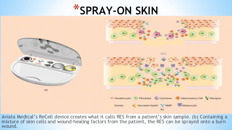
Aviata Medical’s ReCell device creates what it calls RES from
a patient’s skin sample. (b) Containing a mixture of skin cells and wound-healing factors from the patient, the RES can be sprayed onto a burn wound.
Слайд 17While test-tube skin can help when there’s not enough donor skin,
its disadvantage is that it can take weeks to grow the skin cells in the lab. This delay means lengthy hospital stays and increased risk of infection and scarring. A new technology known as spray-on skin promises to speed up the process and reduce risks. Using a small biopsy of a patient’s healthy skin, Avita Medical’s ReCell device (Figure 1) creates what the U.K.-based company calls Regenerative Epithelial Suspension (RES). Within about 30 minutes, the RES, which contains cells and wound-healing factors from the patient, can be sprayed onto the burn wound, covering an area up to 80 times the size of the biopsy site. The system can be used alone to replace the epidermis, but for reconstructing the lower dermis layer in patients with deep burns, it must be used in conjunction with other technology.
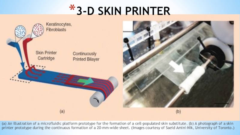
Слайд 18(a) An illustration of a microfluidic platform prototype for the formation
of a cell-populated skin substitute. (b) A photograph of a skin printer prototype during the continuous formation of a 20-mm-wide sheet. (Images courtesy of Saeid Amini-Nik, University of Toronto.)
3-D SKIN PRINTER
Слайд 19The process begins with stem cells that are harvested from a
patient, then turned into specific types of skin cells, and multiplied in the lab. The printer precisely determines the spatial location of each cell, combining them with a gel-like scaffolding substance. The result: strips of substitute skin (Figure 2). “The main thing is that, for burn patients, we need to manufacture a skin substitute that has a dermis component plus an epidermal component,” explains Amini-Nik.
Слайд 21Indeed, one reason burns are particularly difficult to treat is because
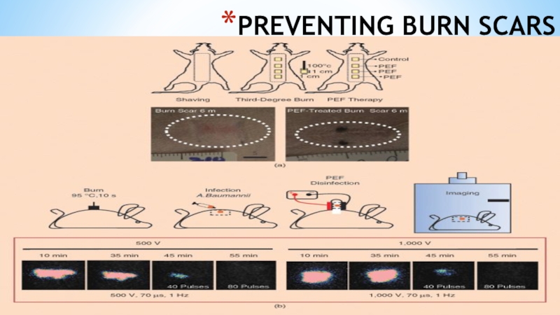
they destroy more than just skin cells. “You are certainly destroying cells, but you are also destroying the extracellular matrix around the cells, the conduits that carry blood via small arterioles, venules or capillaries, and nerves,” explains Martin Yarmush, M.D., Ph.D., who directs the Center for Engineering in Medicine at Massachusetts General Hospital. “Burned tissue is basically overcooked meat.” The technique works because the PEFs create holes in the membranes of cells, allowing the cells’ internal contents to leak out. Importantly, PEF treatment leaves other structures—like the extracellular matrix and the basic organization and structures of sweat glands, arteries, veins, and hair follicles—intact. In fact, Yarmush’s group has observed the regrowth of accessory organs following PEF treatment of normal rat skin, regrowth that could have important clinical applications down the line.
Слайд 22MONITORING HEALING
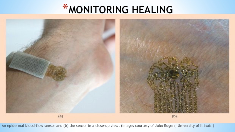
An epidermal blood-flow sensor and (b) the sensor in a
close-up view. (Images courtesy of John Rogers, University of Illinois.)
Слайд 23Flexible electronics are the name of the game for Northwestern University
professor John A. Rogers, Ph.D. His group has developed a series of epidermal electronic sensors [5] that can be used to monitor the health of skin, including the healing of skin wounds following burns or surgery (Figure 4). “These are devices that have kind of the properties of skin,” says Rogers. “They’re super-thin and soft and can—in a nonirritating, noninvasive way—laminate right onto the surface of the skin.” These devices can make a range of different measurements useful for clinicians who want to know how successfully a wound is healing: they can provide information about inflammation and possible infection via high-precision spatial temperature mapping, indicate how hydrated the skin is by measuring thermal conductivity, and measure blood flow to monitor revascularization. “We can do all of those different things in a single platform,” Rogers explains.
